[ Ana Sayfa | Editörler | Danışma Kurulu | Dergi Hakkında | İçindekiler | Arşiv | Yayın Arama | Yazarlara Bilgi | E-Posta ]
Fırat Tıp Dergisi
2017, Cilt 22, Sayı 3, Sayfa(lar) 118-125
[ Özet ] [ PDF ] [ Benzer Makaleler ] [ Yazara E-Posta ] [ Editöre E-Posta ]
Diffusion-Weighted MR Imaging of Acute Abdominal Pain: A Prospective Study of 720 Patients, Assessing the Diagnostic Value of Diffusion-Weighted Imaging Prior Computed Tomography Scanning
Oğuzhan ÖZDEMİR1, Yavuz METİN1, Nurgül ORHAN METİN1, Özlem BİLİR2, Özcan YAVAŞİ2, Ali KÜPELİ3
1Recep Tayyip Erdoğan Üniversitesi Tıp Fakültesi, Radyoloji Anabilim Dalı, Rize, Türkiye
2Recep Tayyip Erdoğan Üniversitesi Tıp Fakültesi, Acil Tıp Anabilim Dalı, Rize, Türkiye
3Muş Devlet Hastanesi, Radyoloji Kliniği, Muş, Türkiye
Keywords: Akut Karın Ağrısı, Bilgisayarlı Tomografi, Difüzyon Ağırlıklı MR Görüntüleme, Acute Abdominal Pain, Computed Tomography, Diffusion-Weighted MR Imaging
Summary
Objective: We aimed to evaluate the contribution of diffusion-weighted magnetic resonance imaging (DW-MRI) to computed tomography (CT) in patients with acute abdominal pain admitted to the emergency department.

Material and Method: Between August 2014 and November 2015, a total of 2365 patients with acute abdominal pain were admitted to the emergency department. 1680 of those underwent CT imaging. In 720 patients (mean age, 50.17; range, 16-89 years: 379 females, 341 males) DW-MRI was performed before CT scans. 960 patients who had no diffusion imaging were excluded from this study. Pregnant women, children under the age of 16 years old, most of urinary stones and all of bowel obstructions were also excluded from this study. The strategy of imaging review was as follows: 1-evaluation of DW-MRI alone, 2-evaluation of CT alone, and 3-evaluation of both modalities (DW-MRI and CT images). Images were evaluated by different radiologists with at least 5 years of experience in abdominal imaging interpretation, blind to the final diagnosis. Every reader was aware of the clinical symptoms and laboratory results of patients.

Results: The sensitivity and accuracy of combined imaging (DW-MRI and CT) was higher than CT alone for the detection of cause in acute abdominal pain. This was dramatically higher in non-enhanced CT (NECT) rather than contrast-enhanced CT (CECT) scanning.

Conclusion: DW-MRI is a noninvasive technique that may be used to improve the accuracy of CT in many cause of acute abdominal pain, especially in patients undergoing non-enhanced CT scans.

  • Top
  • Summary
  • Introduction
  • Methods
  • Results
  • Disscussion
  • References
  • Introduction
    An accurate and fast diagnosis is essential for the appropriate management of acute abdominal pain in the emergency department. It has been shown that emergent abdominal surgical procedures account for approximately 53% of all nontrauma-related surgical interventions performed in the acute care setting1,2. Ultrasonography (US) and computed tomography (CT) are the traditional imaging modalities used for acute abdominal pain in the emergency department. Diffusion-weighted magnetic resonance imaging (DW-MRI) of the abdomen and pelvis has been increasingly used since the 1990s with the development of stronger diffusion gradients, faster imaging sequences, and improvements in technology and magnetic resonance instrumentation3,4. There are many studies with DW-MRI regarding abdominal malignant or inflammatory processes5,6.

    It is well known that DW-MRI relies on the principle of different degrees of mobility of molecules, primarily water molecules, among different tissues at cellular level. Tissue cellularity, cell membrane integrity, types of macromolecules present, perfusion level, and physicochemical properties affect the diffusion of water molecules7. Diffusion is inversely related to cellularity, cell membrane integrity and lipophilicity8,9.

    Restricted diffusion is observed in tissues with high cellularity (tumors, abscesses, fibrosis and cytotoxic edema)8,9. The images are obtained in short interval times and without the requirement of contrast medium10,11.

    To our knowledge, this is the first study that covers many causes of acute abdominal pain to determine the added value of DW-MRI prior to CT scanning.

    We hypothesize that DW-MRI performed before CT will increase the diagnostic accuracy in acute abdominal pain. This could be especially valuable in the assessment of non-enhanced CTs (NECT).

  • Top
  • Summary
  • Introduction
  • Methods
  • Results
  • Disscussion
  • References
  • Methods
    Patient selection and inclusion criteria
    Between August 2014 and November 2015, a total of 2365 patienst with acute abdominal pain were admitted to emergency department of tertiary care training and research hospital. 1680 of those underwent CT ima-ging. Only for 720 patients (42.8%) DW-MRI was performed prior to CT scans. So, 960 patients (57.2%) who had no prior DW-MRI, were excluded from this study. Of the 720 patients, 161 (22.4%) with impaired renal function underwent NECT and 559 (77.6%) had contrast-enhanced CT (CECT). All patients underwent ultrasonography (US) as the initial imaging method. None of them had further imaging when the diagnosis was made at initial US when combined with compatib-le clinical and laboratory findings. Pregnant women, children under the age of 16, Thus, the final study sample consisted of 720 patients [mean age, 50.17; range, 15-89 years: 379 females (52.7%), 341 males (47.3%)]. The study population is shown in Figure. 1.


    Click Here to Zoom
    Figure 1: Patient flow diagram

    All patients who underwent DW-MRI and CT, had acute abdominal pain starting from hours to a few days with variable degrees of symptoms (Table 1).


    Click Here to Zoom
    Table 1: Patient flow diagram

    Approval from the hospital ethics committee and in-formed consents from all patients were obtained.

    Imaging protocols
    DW-MRI examination was performed in total of 720 patients prior to CT scan. All examinations were made on a 1.5-T MRI unit (Magnetom® Aera; Siemens, Erlangen, Germany) with an 18-chanel phased -array body coil. The imaging protocol consisted of an axial diffusion-weighted single-shot echoplanar sequense with fat suppression, without breath holding (TR, 7500 ms; TE, 62-80 ms; matrix, 192x192; slice thickness, 5 mm; gap, 6 mm; FOV, 400 mm; PAT factor, 2; acqusi-tion time, 3 min; b values, 0, 500, and 1000 s/mm2). No extra MR sequences other than diffusion were used in this study.

    A CT scan was obtained after the laboratory results. NECT was performed in 161 patients with impaired renal function, while 559 patients with normal renal function underwent CECT. CT was performed with a 16- slice multidedector-row scanner (Toshiba Alexion™/ Advance, Toshiba Medical Systems Corpo-ration Nashu, Japan). CT was obtained after the initial laboratory results. Those with impaired renal function underwent non-enhanced CT (22.4%). In the remaining patients (77.6%), dynamic images with arterial (scan-ning delay, 20-30 s), portal venous (scanning delay, 60-70s) and equilibrium (scanning delay, 2-3 min) phases were obtained after injection of a total of 100 ml non-ionic contrast material containing iodine con-centration of 300 mg/mL by a power injector at 4 mL/s velocity. None of the patients were given enteral cont-rast medium.

    Image and statistical analysis
    Three radiologist with at least 5 years of experience in abdominal imaging interpretation prospectively re-viewed the examinations. Every reader was aware of the clinical symptoms and laboratory results of pati-ents. Each reader was blind to the final diagnosis. The first reader evaluated DW-MRI images in an indepen-dent workstation (Syngo.via, Siemens). The reader was blinded to CT images. Three b values (0, 500 and 1000 s/mm2) using a respiratory-triggered single-shot echo-planar imaging sequence were used for DW imaging. DW images with highest b value was used, and bright signal was considered to be positive for regions of interest. Apparent diffusion coefficient (ADC) maps were also used to be sure of the pathology. Three diffe-rent ADC values were calculated using region of inte-rest (ROI) placed centrally, and the mean value was taken as the accepted measurement. The second reader interpreted the CT images. The reader was blind of DW images. The result was either negative with nor-mal findings or positive for a specific pathology as a cause of acute abdominal pain.

    The third reader made a diagnosis after reviewing both DW and CT images. The reviewer evaluated the ima-ges in another independent workstation (Syngo. via, Siemens).

    After the blind reviews and combined reviews (DW and CT images), the final diagnosis that we defined as ‘accepted diagnosis’ was made by three radiologists in consensus, on the basis of clinical and laboratory fin-dings. Statistical analysis was done in comparison with ‘accepted diagnosis’.

    Statistical Package for the Social Sciences (SPSS 13.0 Statistical Software, SPSS Inc., Chicago, IL, USA) was used for all statistical analysis. The sensitivity, specifi-city, positive predictive value (PPV) and negative pre-dictive value (NPV), and accuracy were determined on the basis of reviewing DW-MRI only, CT only, and combined images (DW and CT images). The patients were again divided into contrast enhanced CT (CECT) group and non-enhanced CT (NECT) group, and the sensitivity, specificity, positive predictive value (PPV) and negative predictive value (NPV), and accuracy rates were again calculated. The weighted Cohen’s kappa coefficient analysis with a CI of 95% was used to assess the agreement between only DW-MRI and only CT observers. When agreement was perfect, kap-pa and ICC values had a maximum of 1.0; a value of 0 indicated poor agreement. p values of less than 0.05 were accepted to be statistically significant.

  • Top
  • Summary
  • Introduction
  • Methods
  • Results
  • Disscussion
  • References
  • Results
    The results are shown in Table 2. Of the 720 patients, 592 were found to be normal (82.2%) and 128 had various causes of acute abdominal pain (17.8%), as a result of all reviews and the final consensus given by the three radiologists. For those who were found to be radiologically normal, close follow up was decided, and the clinically stable ones were discharged from the emergency department. 23 acute appendicitis (17.9%), 17 acute pyelonephritis (13.2%), 9 acute pancreatitis (7%), 11 acute cholecystitis (7%), 8 acute diverticulitis (6.2%), 6 superior mesenteric vein (SMV) thrombus with intestinal ischemia (4.6%), 5 inflammatory bowel disease (4.6%), and 4 intraabdominal abscesses (3.1%).


    Click Here to Zoom
    Table 2: Results of CT alone, DWI alone, combined images; NECT alone, CECT alone and overall diagnosis

    The statistical analysis related to each single review and combined reviews (DW and CT images) are listed in Table 3. The interobserver agreement was significant with a weighted kappa coefficient of 0.63 (p <0.001). The sensitivity, specificity, and accuracy was found to be 53.6%, 100%, 86.3% for NECT; 59.4%, 100%, 92.9% for CECT; and 53.1%, 100%, 91.6% for total CTs, respectively. It was found to be 94.5%, 100%, and 99%, respectively for DW-MRI. The sensitivity, specificity, and accuracy was 100% for overall combi-ned imaging methods (DW and CT images). The result of the combined methods was the same as final ‘accep-ted diagnoses’ made by the consensus of three radiolo-gists. When used alone, DW-MRI alone failed to diag-nose 10 of the cases, while CT alone failed to diagnose 60 of the cases (Table 3).


    Click Here to Zoom
    Table 3: Results of accepted diagnoses of 128 patients

  • Top
  • Summary
  • Introduction
  • Methods
  • Results
  • Disscussion
  • References
  • Discussion
    US is an easily and widely used imaging modality as the first step tool in the emergency department, especi-ally in the evaluation of children and pregnant patients. But it has many limitations: sonographer dependency, obesity, abdominal gas, and ineffective ability to solve complicated disease processes12,13. Acute abdomi-nal pain in pregnant is another challenging problem that US is generally inefficient to overcome. Lazarus et al.14 has reported that in 30% of pregnant patients with abdominal pain in whom the US study was nega-tive, additional imaging gave important findings, with 64% of these new findings requiring surgical interven-tion. We did not use the findings of US in our study. Pregnant patients and children under 16 years old were also excluded from our study.

    CT is the most commonly used modality in acute ab-dominal pain with high sensitivity and specifity over 90%. However, it is well known that, ionizing radiation and use of contrast material are the disadvantages. This results in an obstacle especially in the evaluation of pregnant patients and children15. It is also stated that ionizing radiation is causing increasing concern, both in the general population and in the medical community16. Although, abdominal CT can be performed without contrast material, it is stated that the intravenous administration of contrast material facilates the evaluation with good accuracy and a high level of di-agnostic confidence, especially in rendering diagnosis in thin patients, in whom fat interfaces may be almost absent17. In our study, we have found that CECT was more sensitive than NECT, with better accuracy rates (92.9% vs 86.3%). Our accuracy rate for all CTs in acute abdominal pain was 91.6%, compatible with the literature, whereas we had a lower sensitivity rate (53.3%) compared to a previous study in which it was reported to be generally over 90%17. This lower rate may be due to exclusion of most urinary emergencies from the study sample, exclusion of all bowel obstruc-tions, high number of complex disease processes and nonenhanced CTs. None of our patients used enteral contrast medium. It seems that lack of rectal contrast does not interfere an accurate diagnosis in acute abdo-minal pain. In a study with a series of 1021 patients, it was found that there were no inconclusive CT scans due to the lack of enteral contrast material18. Studies show that initial US followed by CT examination as a diagnostic strategy regarding acute abdominal pain, reduces unnecessary CT scans, and thus reducing ra-diation exposure19,20. All of our patients un-derwent initial US examination, followed by DW-MRI and CT scan. DW-MRI and CT was decided when US was either inefficient or a further diagnostic modality was needed on the basis of clinical and laboratory evaluation. In our study, in patients who were found to have acute appendicitis at initial US, CT was not per-formed, and these patients were excluded from the study. This was also applicable for patients who were diagnosed as acute cholecystitis at initial US. DW-MRI and CT imaging were obtained in these patients when the clinical picture and laboratory results (e.g. high liver function tests and amylase levels) pointed cholan-gitis or pancreatitis, as in 22 patients in our study. Ele-ven of them were found to be cholecystitis, 2 cholecys-titis with cholangitis, and 9 pancreatitis. Patients with hydroureteronephrosis at initial US had NECT without DW-MRI, and therefore they were excluded from the study. We had a total of 17 patients with acute pyelo-nephritis. Initial US evaluation of these patients did not show prominent degree of hydroureteronephrosis or give enough information regarding the clinical status, so DW-MRI and CT imaging were obtained.

    There are also many attempts to decrease the radiation dose in CT scans and many studies have been publis-hed regarding this issue, so far17. Exposure to ioni-zing radiation is a disadvantage of CT. The dose of radiation associated with abdominal CT in acute abdo-men is approximately 10 mSv. It is estimated that, for a 25 year old patient, the risk of cancer induction for such a CT scan is about 1 in 900, the risk of fatal can-cer induction is 1 in 1800; for a 50 year old, the equivalent risks are 1 in 1500 and 1 in 2500, respectively21,22. In general, consensus exists that the information obtained with diagnostic CT outweighs the risk associ-ated with radiation and that the risk of cancer induction should be seen in the light of the lifetime cancer risk18.

    There are many studies in the literature reporting the use of MRI for management of acute abdominopelvic pain. Recent advances in MRI hardware and software have allowed the development of rapid imaging tech-niques that are particularly applicable for emergency department indications23-25. Although CT scanning is the primary imaging method used in the emergency department, MRI is increasingly being used for acute abdominal pain. Being free of ionizing radiation and no need of iodinated contrast medium are the advantages of MRI. Furthermore, a majority of acute abdominopelvic diseases don’t require the use of intravenous contrast medium1,17,24-26. Abdominal DW-MRI has been increasingly used with improvements in tech-nology and MRI instrumentation since 1990s. It is well known that DW-MRI relies on the principle of diffe-rent degrees of mobility of molecules, primarily water molecules, among different tissues at cellular level. Tissue cellularity, cell membrane integrity, types of macromolecules present, perfusion level, and physi-cochemical properties affect the diffusion of water molecules1,25. Diffusion is inversely related to cellularity, cell membrane integrity and lipophilicity. Restricted diffusion is observed in tissues with high cellularity (tumors, abscesses, fibrosis and cytotoxic edema)6,27-29. The images are obtained in short interval times and without the need of contrast me-dium. Quantitative analysis may be performed with the generation ADC maps from diffusion images obtained at different b values27. Although at least two b va-lues are required for DW imaging analysis, it is stated that the application of a greater number of b values will improve the accuracy of the calculated ADC30 In our study, we used 0, 500, and 1000 s/mm2 standard b values. We did not use any contrast enhanced MRI or other MRI sequenses, in order to gain time for urgent cases.

    Interestingly, our study revealed a high sensitivity, specificity, and accuracy rates for DW-MRI (94.5%, 100%, 99%, respectively) which were even higher in combined methods (100%). To our knowledge, this is the first study that covers many causes of acute abdo-minal pain with a combination of DW-MRI and CT imaging. We think that this high rates is the result of combined imaging methods. And also it is a remarkab-le point that, the final diagnosis is made by the consen-sus of three radiologists who also take account of a good clinical and laboratory based evaluation.

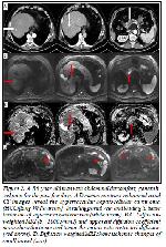
    In our study, DW-MRI had also improved the diagno-sis of complex disease processes. One of them was tumoral trombus of SMV caused by hepatocellular cancer (HCC) invasion (Figure. 2).


    Click Here to Zoom
    Figure 2: A. 86-year-old man with abdominal discomfort, generali-zed pain for the past few days. A Dynamic contrast-enhanced axial CT images reveal the hypervascular hepatocellular carcinoma (HCC)[long White arrow], invading portal vein and leading to tumor thrombus of superior mesenteric vein (white arrow). B,C. Diffusion-weighted MRI (b= 1000 s/mm2) and apparent diffusion coefficient maps show the tumor and tumor thrombus with restricted diffusion (red arrow). D. Diffusion-weighted MRI shows ischemic changes of small bowel (star).

    CT successfully revealed the hypervascular liver tumor invading portal vein, reaching to SMV. DW-MRI both showed the malignant nature and extent of invasion as well as ischemic changes of small bowel, thus contri-buting to the diagnosis. At CT the ischemia of small bowel was occult. DW-MRI also improved the visuali-zation of the tumoral thrombus.

    Our study also showed that CT was inefficient to cha-racterize pyelonephritis, especially with NECT. Two of 17 patients with pyelonephritis could be defined by CT-only reader, while 16 were diagnosed by DW-MRI-only reader, and all were diagnosed with the combination of examinations.

    We have also found that in 14 intestinal ischemia with different causes, CT-only reader could show ischemic changes in 3 of them, and DW-MRI-only reader could characterize all cases of ischemia. With combined reviews, both the cause and ischemic change of intesti-ne were clearly visualized.

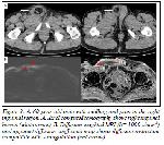
    Similarly, in the evaluation of stranguled inguinal her-nias, DW-MRI reader diagnosed all the strangulations accompanying the inguinal hernias (Figure 3).


    Click Here to Zoom
    Figure 3: A. 60-year-old man with swelling and pain in the right inguinal region. A. Axial computed tomography shows right inguinal hernia (white arrow). B. Diffusion-weighted MRI (b= 1000 s/mm2) and apparent diffusion coefficient map shows diffusion restriction compatible with strangulation (red arrow).

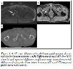
    We had an interesting case with right lower quadrant pain that was found to be due to right pubic bone metastasis visualized on DW-MRI, which was nonvisible at CT (Figure. 4).


    Click Here to Zoom
    Figure 4: A. 67-year-old man with right lower quadrant pain due to right pubic bone metastasis. (A,B) Diffusionweighted MRI (b=1000 s/mm2) and aparent diffusion coefficient map showed restricted diffusion of right pubic bone (white arrow) and C axial CT images of pubic bone was normal.

    Relatively small number of some sample emergencies, especially those with urinary stones that is one of the most common cause of acute abdominal pain, exclu-sion of bowel obstructions, lack of a gold standart comparison method other then the final diagnosis as we called ‘accepted diagnosis’ made by decision of three radiologists, and low spatial resolution of DW-MRI were our major limitations.

    In conclusion, DW-MRI is a non-invasive imaging method which has no ionizing radiation, dose not requ-ire contrast media, and can be easily performed in a short time. We suggest that DW-MRI should be added to the imaging protocol for acute abdominal pain in emergency departments, especially for pregnant woman and children. DW-MRI may aid in the detec-tion of the acute focus (inflammation or infection) with its bright signal, and thus guiding a prompt diagnosis. We propose that this is especially critical when a NECT scan is planned.

    DW-MRI can also have a role in monitoring patients with acute abdominal pain, who are not operated, and need a close follow up. So it can help to prevent un-necessary CTs in follow p, and thus reducing exposure of ionizing radiation.

    Ethical approval: All procedures performed in studies involving human participants were in accordance with the ethical standards of the institutional and/or national research committee and with the 1964 Helsinki decla-ration and its later amendments or comparable ethical standards.

  • Top
  • Summary
  • Introduction
  • Methods
  • Results
  • Discussion
  • References
  • References

    1) Singh A, Danrad R, Hahn PF, Blake MA, Muel-ler PR, Novelline RA. MR imaging of the acute abdomen and pelvis: Acute appendicitis and be-yond. Radiographics 2007; 27: 1419-31.

    2) Pedross I, Rofsky NM. MR imaging in abdomi-nal emergencies. Radiol Clin North Am 2003; 41: 1243-73.

    3) Qayyum A. Diffusion-weighted imaging in the abdomen and pelvis: concepts and applications. Radiographics 2009; 29: 1797-1810.

    4) Bayraktutan U, Oral A, Kantarci M, et al. Diag-nostic performance of diffusion-weighted MR imaging in detecting acute appendicitis in child-ren: comparison with conventional MRI and surgical findings. J Magn Reson Imaging 2014; 39: 1518-24.

    5) Onur MR, Poyraz AK, Kocakoc E, et al. Diag-nosis of peritoneal metastases with abdominal malignancies: role of ADC measurement on dif-fusion weighted MRI. Eurasian J Med 2012; 44: 163-8.

    6) Koh DM, Collins DJ. Diffusion-weighted MRI in body: application and challenges in oncology. AJR Am J Roentgenol 2007; 188: 1622-35.

    7) Islim F, Salik AE, Bayramoglu S, Guven K, Alis H, Turhan AN. Non-invasive detection of infection in acute pancreatic and acute necrotic collections with diffusion-weighted magnetic resonance imaging: preliminary findings. Ab-dom Imaging 2014; 39: 472-81.

    8) Kele PG, van der Jagt EJ. Diffusion weighted imaging in the liver. World J Gastroenterol 2010; 16: 1567-76.

    9) Bittencourt LK, Matos C, Coutinho AC. Diffu-sion-weighted magnetic resonance imaging in the upper abdomen: technical issues and clinical applications. Magn Reson Imaging Clin N Am 2011; 19: 111-31.

    10) Kiryu S, Dodanuki K, Takao H, et al. Free-breathing diffusion-weighted imaging for the assessment of inflammatory activity in Crohn’s disease. J Magn Reson Imaging 2009; 29: 880-6.

    11) Oto A, Schmid-Tannwald C, Agrawal G, et al. Diffusion-weighted MR imaging of abdomino-pelvic abscesses. Emerg Radiol 2011; 18: 515-24.

    12) Curtin KR, Fitzgerald SW, Memcek AA, Hoff FL, et al. CT diagnosis of acute appendicitis: imaging findings. AJR Am J Roentgenol 1995; 64: 905-9.

    13) Friedland JA, Siege MIJ. CT appearance of acute appendicitis in childhood. AJR Am J Ro-entgenol 1997; 168: 439-42.

    14) Lazarus E, Mayo-Smith WW, Mainiero MB, Spencer PK. CT in the evaluation of nontrauma-tic abdominal pain in pregnant woman. Radio-logy 2007; 244: 784-90.

    15) Thirumoorthi AS, Fefferman NR, Ginsburg HB, et al. Managing radiation exposure in children-reexamining the role of ultrasound in the diag-nosis of appendicitis. J Pediatr Surg 2012; 47: 2268-72.

    16) Lubarsky M, Kalb B, Sharma P, Keim SM, Martin DR. MR imaging for acute non-traumatic abdominopelvic pain: rational and practical considerations. Radiographics 2013; 33: 313-37.

    17) Stoker J, van Randen A, Laméris W, et al. Ima-ging patients with acute abdominal pain. Radio-logy 2009; 253: 31-46.

    18) Laméris W, van Randen A, van Es HW, et al. Imaging strategies for detection of urgent condi-tions in patients with acute abdominal pain: di-agnostic accuracy study. BMJ 2009; 338: b2431.

    19) Ng CS, Watson CJ, Palmer CR, et al. Evalua-tion of early abdominopelvic computed tomog-raphy in patients with acute abdominal pain of unknown cause: prospective randomised study. BMJ 2002; 325: 1387.

    20) Sala E, Watson CJ, Beadsmoore C, et al. A randomized, controlled trial of routine early ab-dominal computed tomography in patients pre-senting with non-specific acute abdominal pain. Clin Radiol 2007; 62: 961-9.

    21) The 2007 recommendations of the international commission on radiological protection: ICRP publication 103. Ann ICRP 2007; 37: 1-332.

    22) Board on radiation effects research (BRER). Health risks from exposure to low levels of io-nizing radiation: BEIR VII phase 2. Washington DC: National Academics Press, 2006.

    23) Katz DS, Klein MA, Ganson G, Hines JJ. Ima-ging of abdominal pain in pregnancy. Radiol Clin North Am 2012; 50: 149-71.

    24) Spalluto LB, Woodfield CA, DeBenedectis CM, Lazarus E. MR imaging evaluation of abdomi-nal pain during pregnancy: appendicitis and ot-her nonobstetric causes. Radiographics 2012; 32: 317-34.

    25) Leyendecker JR, Gorengaut V, Brown JJ. MR imaging of maternal diseases of the abdomen and pelvis during pregnancy and the immediate postpartum period. Radiographics 2004; 24: 1301-16.

    26) Kalb B, Sharma P, Salman K, Ogan K, Pattaras JG, Martin DR. Acute abdominal pain: is there a potential role for MRI in the setting of the emergency department in a patient with renal calculi? J Magn Reson Imaging 2010; 32: 1012-23.

    27) Thoeny HC, De Keyzer F. Extracranial applica-tions of diffusion-weighted magnetic resonance imaging. Eur Radiol 2007; 17: 1385-93.

    28) Chan JH, Tsui EY, Luk SH, Fung AS, Yuen MK, Szeto ML, Cheung YK, Wong KP. Diffu-sion weighted MR imaging of the liver: distin-guishing hepatic abscess from cystic or necrotic tumor. Abdom Imaging 2001; 26: 161-5.

    29) Holzapfel K, Eiber MJ, Fingerle AA, Bruegel M, Rummeny EJ, Gaa J. Detection, classifica-tion, and characterization of focal liver lesions: value of diffusion-weighted MR imaging, gadoxetic acidenhanced MR imaging and the combination of both methods. Abdom Imaging 2012; 37: 74-82.

    30) Pagani E, Bizzi A, Di Salle F, De Stefano N, Filippi M. Basic concepts of advanced MRI techniques. Neurol Sci 2008; 29: 290-5.

  • Top
  • Summary
  • Introduction
  • Methods
  • Results
  • Discussion
  • References
  • [ Başa Dön ] [ Özet ] [ PDF ] [ Benzer Makaleler ] [ Yazara E-Posta ] [ Editöre E-Posta ]
    [ Ana Sayfa | Editörler | Danışma Kurulu | Dergi Hakkında | İçindekiler | Arşiv | Yayın Arama | Yazarlara Bilgi | E-Posta ]