 |
 |
| [ Ana Sayfa | Editörler | Danışma Kurulu | Dergi Hakkında | İçindekiler | Arşiv | Yayın Arama | Yazarlara Bilgi | E-Posta ] | |
| Fırat Tıp Dergisi | |||||
| 2017, Cilt 22, Sayı 4, Sayfa(lar) 212-215 | |||||
| [ Özet ] [ PDF ] [ Benzer Makaleler ] [ Yazara E-Posta ] [ Editöre E-Posta ] | |||||
| Anesthetic Management of a Patient with Frontometaphyseal Dysplasia (Gorlin-Cohen Syndrome) Undergoing Genu Recurvatum Correction Surgery | |||||
| Mehmet CANTÜRK | |||||
| Ahi Evran University Education and Research Hospital, Anesthesiology and Reanimation, Kırşehir, Turkey | |||||
| Keywords: Frontometafizyal Displazi, Genel Anestezi, Havayolu Yönetimi, Genu Rekurvatum, Frontometaphyseal Dysplasia, General Anesthesia, Airway Management, Genu Recurvatum | |||||
| Summary | |||||
Frontometaphyseal dysplasia (FMD) is a rare X-linked hereditary disorder. Skeletal deformities are common manifestations. Patients usually undergo surgeries early in life. The anesthetic management of a patient with FMD is presented in the current case report. A 6 years old female patient, weighing 20kg and height 120cm, admitted to hospital and scheduled for genu recurvatum correction. Her eyebrows were prominent, she had defects in teeth, proptosis, micrognathia, pectus carinatus, genu recurvatum and bowing tibia, and 2o over 6o murmur. Right tympanic membrane was perforated. Induction and maintenance of anesthesia was uneventful. FMD patients are candidates for difficult airway management and anesthesiologists taking care of a patient with FMD must be prepared for an anticipated difficult airway. For a safe anesthetic management in FMD, a careful and detailed preoperative visit and consultation with related departments, strict monitorization at the operation room and being prepared for an anticipated difficult airway is essential. |
|||||
| Introduction | |||||
Frontometaphyseal dysplasia (FMD), also known as Gorlin-Cohen syndrome is an X-linked hereditary disorder 1. Up to date, approximately 100 cases have been described on Medline. Data regarding anesthetic management of FMD is limited. FMD is now known to have an X-linked dominant hereditary inheritance. The responsible gene is Xq28 which has a role in encoding filamin A 10. The characteristic findings of FMD include craniofacial abnormalities, skeletal abnormalities, hearing problems, and wasting of extremity muscles. Besides these skeletal deformities, congenital heart malformations, subglottic stenosis, asthenia, muscular underdevelopment, hearing loss or deficits, and in male patients urinary symptoms may also coincide. Scoliosis and hearing loss, either sensory and/or conductive, develop progressively 3,4. Skeletal manifestations gain extreme importance for anesthesiologists taking care of patients with FMD. Coarse face shape, wide nasal bridge, incomplete sinus development, partial anodontia, delayed and/or defective tooth eruption, arched palate, small mandible, and subglottic stenosis are characteristic findings in FMD which of all are independent risk factors for airway management 3-5. Respiratory difficulties, subglottic stenosis, chest wall deformities, underdeveloped musculature, and congenital heart diseases are predisposing risk factors for safe anesthesia management and postoperative care. Patients with FMD syndrome need surgery at an early phase of their lives due to the musculoskeletal deformities. To provide an uneventful anesthesia management, preoperative assessment of the patient, informing both the patient and the family about the interventions being planned at the operation room and postoperative care is very important. In this case report anesthetic management of a patient with FMD syndrome is presented. |
|||||
| Case Presentation | |||||
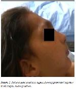
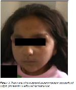
A 6 year old, 20kg weighing, 120cm tall female patient with known FMD syndrome for three years was scheduled for correction of genu recurvatum deformity after the written informed consent was obtained from her parents. The patient had prominent supraorbital ridges, prominent cheeks, and malocclusion of the teeth, proptosis, micrognathia, pectus carinatus, genu recurvatum, and bowing tibia (Figure 1 and Figure 2).
On her physical examination; auscultation of the lungs were normal, there was 2o over 6o heart murmur on mitral focus. She had pectus carinatus deformity, arched palate, hypertelorism, wide nasal bridge, defective teething, and abnormal external ear anatomy. Biochemistry results, total blood count results and ECG were in physiological range. Right tympanic membrane was perforated. There was hypertropia on the left eye. Her radiologic findings revealed bowing of the 12th rib, small mandibles, arched palate, prominent supraorbital ridges, aplastic frontal sinuses, stenotic iliac bone, coxa valgus deformity, and bowing of the tibia. The patient was classified as Mallampati score of III and Cormack Lehane score of III. At the preoperative visit, both the patient and her parents were informed about FMD, the course of the disease, the anesthetic and surgical interventions being planned, the anticipated postoperative course, any probable side effects and their treatments. After obtaining verbal and written informed consent from the parents an intravenous line was secured on dorsum of left hand. Midazolam 1mg was infused slowly for premedication. The anesthetic preparation for an anticipated difficult airway management and intubation constituted of transparent face masks, a laryngoscope with both Miller and Macintosh blades, cuffed intubation tubes between 3# to 5#, stylets, flexible laryngoscope, video laryngoscope, percutaneous tracheostomy set, and surgical suction as defined by the guidelines of American Society of Anesthesiology. Emergency medication as atropine, adrenaline and sugammadex for reversal of muscle relaxation was ready for use in case of emergency. An ENT specialist was prepared in sterile coats in case of a need for emergency tracheostomy. Upon arrival to the operating room, the patient was monitorized with standard ASA monitors (electrocardiogram, percutaneous pulse oximeter, non invasive blood pressure and end tidal carbon dioxide attached to anesthesia circuit). The basal heart rate was 120 beats/min and blood pressure 100/60 mmHg. After preoxygenation for 3 minutes, the induction of anesthesia was achieved by intravenous 1mg/kg lidocaine, 6mg/kg thiopental and 1mg/kg rocuronium. Following mask ventilation for two minutes, intubation was achi-eved with #2 Macintosh blade and #4.5 cuffed endotracheal tube. The proper localization of endotracheal tube was confirmed by auscultation and capnograph. Anesthesia was maintained with 2% sevoflurane in 50% O2/N2O mixture for 3 hours lasting operation. When spontaneous ventilation was adequate and patient fully recovered, she was uneventfully extubated and discharged to the recovery room. During the follow up period in recovery room she did not manifest any problem in ventilation and discharged to ward after four hours of follow up. |
|||||
| Discussion | |||||
Frontometaphyseal dysplasia also known as Gorlin-Cohen syndrome is a rare member of craniotubular dysplasia family 1. It is accepted to be an X-linked hereditary syndrome 2. There is a functional mutation in Xq28 locus which is responsible for encoding filamin. The common clinical findings of the syndrome includes prominent supraorbital ridges, defects in teeth and malocclusion, micrognathia, congenital heart diseases, restrictive type pulmonary diseases, subglottic stenosis, hearing loss and other structural bone deformities. 3,4 The presence of micrognathia, malocclusion and defective teeth may lead to airway management problems. Takahashi 5 reported a difficult intubation in a FMD patient which was intubated at sitting position with fiberoptic bronchoscope assistance. Since the difficulty in airway management in FMD syndrome is anticipated, difficult airway devices must be set ready in the operating room. Today, the presence of supraglottic airway devices, stylets, flexible laryngoscope blades, video laryngoscopes and fiberoptic bronchoscopes together with a perfect preanesthetic evaluation and physical examination permits the anesthesiologists feel safe when dealing with patients having anticipated difficult airway. Genigara et al. 6 intubated a patient using a combination of dexmedetomidine and ketamine while preserving the spontaneous ventilation. In cases where difficult airway is anticipated, intubation under adequate sedation with preserving spontaneous ventilation is an authoritative choice and can be preferred safely. Since congenital heart diseases commonly coincides FMD syndrome, the patients should be monitorized on arrival to the operation room and must be closely followed-up. Mehta and Schou 4 reported that their patient suffered cardiac arrest at induction with halot-hane. The patient responded to resuscitation in a few seconds and they intubated the patient uneventfully thereafter. The clinical experience of this case is volatile anesthetic agents (especially halothane) is not an appropriate induction agent for patients with FMD syndrome and intravenous induction agents should be preferred. Horasanlı et al. 7 have reported that they have inducted anesthesia with 30 mg propofol and 10 mg of succinylcholine in a case with anticipated difficult airway. They argued that succinylcholine provides a fast muscle relaxation and intubation. In the current case, rocuronium is used for muscle relaxation since it also provides fast repeated intubation and most importantly it does not bare the risk of malignant hyperthermia which is a potential and lethal side effect which may be triggered with succinylcholine use. Sugammadex is a novel agent for reversal of rocuronium and vecuronium induced neuromuscular block, provides very fast reversal approximately in three minutes 8. Presence of such an effective reversal agent for rocuronium favours the use of rocuronium at an anticipated difficult intubation setting. Optimal preanesthetic evaluation, preparing the operation room for anticipated difficult airway management as defined in ASA guidelines 9, close follow up and monitorization, and sufficient preoxygenation before induction of anesthesia makes it safe to use thiopental and rocuronium for induction of general anesthesia. Sufficient muscle relaxation preserves a better view for conventional laryngoscopy. In our case, although all difficult airway devices were set ready, we could manage an uneventful intubation. Algorithmic management steps must be followed up starting from the basic (conventional laryngoscopy) to the last step (tracheostomy) in the management of anticipated difficult airway. The anesthetic management of patients with FMD syndrome requires a careful preanesthetic assessment, physical examination and consultation with related departments, evaluation of the airway, closed follow up and monitorization at operation room and setting the difficult airway devices ready for use; all of which will preserve for the safety of the patient. Conflicts of Interest: Author declares there is no conflict of interest for the case report. Financial Support: Author declares no financial support fort the case report. |
|||||
| References | |||||
1) Gorlin RJ, Cohen MM. Frontometaphyseal dysplasia, a new syndrome. Am J Dis Child 1969; 118: 487-94.
2) Abuelo D N, Erlich O, Schwartz A, Feingold M. picture of the month. Frontometaphyseal dysplasia. Am J Dis Child 1983; 137: 1017-8.
3) Robertson SP. Otopalatodigital syndrome spectrum disorders: Otopalatodigital syndrome types 1 and 2, frontometaphyseal dysplasia and Melnick–Needle syndrome. Eur J Hum Genet 2007; 15: 3-9.
4) Mehta Y, Schou H. The anaesthetic management of an infant with frontometaphyseal dysplasia (Gorlin–Cohen syndrome). Acta Anesthesiol Scand 1988; 49: 957-64.
5) Takahashi K, Kuwahara T, Tanigawara T, Hattori T, Masuno M, Kondo N. Frontome-taphyseal Dysplasia: Patient with ruptured aneurysm of the aortic sinus of Valsalva and cerebral aneurysms. Am J Med Gen 2002; 108: 249-51.
6) Ganigara A, Nishtala M, Chandrika YRV, Chandrakala KR. Airway management of a child with frontometaphyseal dysplasia (Gorlin-Cohen syndrome). J Anesthesiol Clin Pharmacol 2014; 30: 279-80.
7) Horasanlı E, Ornek D, Canturk M, Ozdogan L, Sahin F, Dikmen B. Difficult airway due to protruding macroglossia in a child with lymphangioma. B-ENT 2010; 6: 219-22.
8) Thompson CA. Sugammadex approved to reverse NMBA effects. Am J Health Syst Pharm 2016; 7: 100.
|
|||||
| [ Başa Dön ] [ Özet ] [ PDF ] [ Benzer Makaleler ] [ Yazara E-Posta ] [ Editöre E-Posta ] | |||||
| [ Ana Sayfa | Editörler | Danışma Kurulu | Dergi Hakkında | İçindekiler | Arşiv | Yayın Arama | Yazarlara Bilgi | E-Posta ] |

