[ Ana Sayfa | Editörler | Danışma Kurulu | Dergi Hakkında | İçindekiler | Arşiv | Yayın Arama | Yazarlara Bilgi | E-Posta ]
Fırat Tıp Dergisi
2018, Cilt 23, Sayı 1, Sayfa(lar) 032-035
[ Özet ] [ PDF ] [ Benzer Makaleler ] [ Yazara E-Posta ] [ Editöre E-Posta ]
Multi Sistem Atrofili Hastalarda Tanısal Zorluklar
Murat GÜLTEKİN
Erciyes Üniversitesi Tıp Fakültesi, Nöroloji Anabilim Dalı, Kayseri, Türkiye
Anahtar Kelimeler: Multi Sistem Atrofi, Ataksi, Parkinsonizm, Ayırıcı Tanı, Multiple System Atrophy, Ataxia, Parkinsonism, Differential Diagnosis
Özet
Amaç: Multi sistem atrofi (MSA); parkinsonizm, serebellar ataksi ve otonomik yetmezlik semptomlarından oluşan bir kombinasyon ile karakterize nörodejeneratif bir hastalıktır. Bu çalışmanın amacı; muhtemel MSA tanısı almış hastaların klinik özelliklerini değerlendirmek ve MSA'nın tanısal zorluklarını literatür eşliğinde gözden geçirmektir.

Gereç ve Yöntem: Çalışmaya MSA’nın ikinci konsensüs teşhis kriterlerine göre muhtemel MSA tanısı ile takip edilen 11 hasta dahil edildi. Hasta dosyaları retrospektif olarak incelendi ve en son klinik muayene özellikleri not edildi.

Bulgular: Onbir MSA hastasının yaş ortalaması 65.36 (±5.32) bulundu. Altı hasta kadın cinsiyetteydi. Ortalama hastalık süreleri 4 yıldı. On hasta semptomları başladığı zaman ilk başvurduğu klinisyen tarafından MSA tanısı almamıştı. Sadece bir hasta doğru tanı olarak MSA tanısı almıştı. On hastanın yedisi Parkinson Hastalığı tanısı almışken diğer üç hasta ataksi tanısı almıştı.

Sonuç: Çoğu MSA hastasının ayırıcı tanıdaki güçlüklerden dolayı yaşam boyu doğru tanı alması geç olmaktadır. Bu problemin yeniden düzenlenecek güncel teşhis kriterlerine göre tartışılması gerekmektedir.

  • Başa Dön
  • Özet
  • Giriş
  • Materyal ve Metot
  • Bulgular
  • Tartışma
  • Sonuç
  • Kaynaklar
  • Giriş
    Multiple system atrophy (MSA) is a sporadic neuro-degenerative disease with adult onset characterized by a combination of parkinsonism, cerebellar ataxia, py-ramidal signs, and autonomic deficiency symptoms 1. MSA is one of the Parkinson plus syndromes included in the differential diagnosis of Parkinson's Disease (PD). Its incidence is 4.4/100000 2. There are no therapeutic options which cure the disease apart from symptomatic drugs 3. The second consensus state-ment on the diagnosis of MSA criteria are used for the diagnosis of MSA. According to these criteria, patients are diagnosed with definite, probable, or possible MSA 4.

    In clinical practice, clinicians have to rely on diagnos-tic criteria for MSA. But, there are no reliable biologi-cal markers. Thus, clinicians can need to see brain imaging characteristics of MSA. However, the clinical diagnostic accuracy of MSA varies between 60% and 90% even in the late stage of the disease 5,6. Espe-cially the early stage diagnostic accuracy of MSA is unknown 7. For this reason, there should be im-provements in clinical diagnosis criteria to increase diagnostic accuracy are of great importance.

    The purpose of this study is to review diagnostic chal-lenges of MSA in view of the literature and assess clinical characteristics of patients diagnosed with prob-able MSA with regard to clinical subtypes of MSA-P and MSA-C.

  • Başa Dön
  • Özet
  • Giriş
  • Materyal ve Metot
  • Bulgular
  • Tartışma
  • Sonuç
  • Kaynaklar
  • Materyal ve Metot
    Patients followed-up with MSA diagnosis at the Movement Disorders Polyclinic, Neurology Department, Erciyes University Faculty of Medicine Hospital between June 2014 and December 2016 were included in this study. The approval of the local ethics commit-tee was obtained and the consent of the patients was received. Eleven patients followed-up with probable MSA diagnosis according to the second consensus statement on the diagnosis of MSA criteria were in-cluded in the study 4. The patients were categorized for different types of MSA-P and MSA-C by a clini-cian with experience in movement disorders.

    Patient files were reviewed retrospectively and the latest clinical examination characteristics were noted. Cranial magnetic resonance images (MRI) of all pa-tients were reviewed as well. Characteristics of peri-putaminal hyperintensity (rim sign), putaminal hy-pointensity, atrophy of the brainstem and middle cere-bellar peduncle, and the hot cross bun sign were deter-mined, if present. Additionally; initial symptoms, ini-tial diagnosis, treatment responses, the course of the disease in time, additional symptoms, previous cranial images (if available), disabling symptoms, and pre-dominant clinical characteristics of the patients were assessed with regard to their fit with respective sub-types of MSA. Global disability status was determined for each patient according to their Unified Multiple System Atrophy Rating Scale (UMSARS) scores in order to assess clinical progression of MSA 8.

    Statistics
    Demographical and clinical data obtained from the participants within the scope of the study were evaluat-ed with descriptive statistics. Mean ± SD and median 25 and 75 percentile values are presented. The results of the frequency analysis applied to certain data can be seen in Table 1 and 2. Statistical Package for the Social Sciences 15.0 (SPSS 15.0) was used for analysis.

  • Başa Dön
  • Özet
  • Giriş
  • Materyal ve Metot
  • Bulgular
  • Tartışma
  • Sonuç
  • Kaynaklar
  • Bulgular
    The mean age of the patients was found 65.36. Six patients were female. The average disease time was 4 years. Ten patients were not diagnosed with MSA by the clinician when they applied with the onset of symp-toms. Only one patient was diagnosed with MSA cor-rectly. Seven out of ten patients were diagnosed with PD, whereas the other three patients were diagnosed with ataxia. Table 1 shows the demographic data about the patients.


    Büyütmek İçin Tıklayın
    Table 1: Demographic features of patients with MSA.

    Clinical examination characteristics and observations of the patients were reviewed. Considering initial symptoms, five patients presented cerebellar findings, four patients presented parkinsonian findings, and two patients had cerebellar+parkinsonian findings (mixed). It was found that all patients had atrophy of the brain-stem and middle cerebellar peduncle and the hot cross bun sign. Neurogenic orthostatic hypotension symp-toms were observed in 10 patients. Seven patients were diagnosed with MSA-C and four patients were diag-nosed with probable MSA-P after assessment accord-ing to diagnostic criteria. All MSA-P patients and four MSA-C patients had received L-dopa treatment. How-ever, three MSA-C patients had been forced to give up the L-dopa treatment since it worsened neurogenic orthostatic hypotension symptoms. Clinical character-istics of the MSA patients can be seen in Table 2.


    Büyütmek İçin Tıklayın
    Table 2: The clinical features of patients with MSA.

    Nine out of eleven patients had presented Rapid eye movement (REM) sleep behavior disorder (RBD) symptoms before the onset of symptoms. An assess-ment of the patients based on their latest examination findings showed that all patients required help with balance in everyday life activities according to the global disability scale. Four patients in particular had become wheelchair-bound due to cerebellar symptoms (stage 5). There is seen MRI image of a patient fol-lowed-up with MSA-C below (Figure 1).


    Büyütmek İçin Tıklayın
    Figure 1: The MRI scan of one patient with MSA-C. The hot cross bun sign of the pon and cerebellar atrophy is very common on course of the MSA.

  • Başa Dön
  • Özet
  • Giriş
  • Materyal ve Metot
  • Bulgular
  • Tartışma
  • Sonuç
  • Kaynaklar
  • Tartışma
    In this study, we found that MSA patients are not accu-rately diagnosed especially in initial years where the symptoms start to manifest themselves. Only one in eleven patients followed-up had been accurately diag-nosed. This finding shows that there are diagnostic challenges throughout the clinical course of MSA, especially for patients who are followed-up in places other than centers specializing in movement disorders. The accurate diagnosis of MSA is significant for ap-propriate patient management, particularly in early stages of the disease where the symptoms are not yet fully manifested. However, as far as early diagnosis goes, it is obvious that the existing clinical diagnostic criteria have certain challenges. In this context, it should be mentioned that up-to-date laboratory and clinical findings may contribute to the accuracy of the diagnosis 9.

    Today, an increasing number of publications highlight the challenges in MSA diagnosis 9,10. Existing clin-ical diagnostic criteria used for MSA have various limitations. First of all, these criteria mostly focus on motor findings associated with the disease. Parkinson-ism and cerebellar ataxia non-responsive to L-dopa are not uncommon cases and may be observed in many disorders other than MSA 11,12. Secondly, auto-nomic deficiency and genitourinary dysfunction in particular are not specific to MSA. These may be ob-served in other neurodegenerative diseases and even in healthy individuals between the ages of 50-60 13,14. Thirdly, orthostatic hypotension may be seen in many cases other than MSA. For example, it is reported in a study that 20% of PD patients present orthostatic hypo-tension 15. The fourth limitation is that both auto-nomic deficiency and motor symptoms are necessary for MSA diagnosis. However, autonomic deficiency and motor symptoms do not develop concurrently in many MSA patients. While some patients present auto-nomic deficiency from the onset of the disease, some do not show any symptoms even after 15 years 16. Indeed, one of our patients (Table 2, case 11) had the symptoms for four years, yet did not present orthostatic hypotension to this day.

    A considerable number of patients are not correctly diagnosed with MSA during their lifetime due to diffi-culties in differentiating it from other diseases. (PD, pure autonomic failure, other rare movement disor-ders). MSA may be accompanied by Parkinsonian symptoms quite frequently. About 10% of patients diagnosed with Parkinson’s Disease are reported to have MSA at autopsy. About 29-33% of patients who present isolated late-onset cerebellar ataxia and 8-10% of patients with Parkinsonism develop MSA. For this reason, it is safe to assume a higher prevalence than estimations 17.

    The EMSA registry study examined 437 patients diag-nosed with MSA. Sixty-eight percent of these patients were found to have symptoms that fit MSA-P and 32% were found to have symptoms that fit MSA-C. While 99% of the patients had autonomic deficiency symp-toms (urinary symptoms, orthostatic hypotension, and chronic constipation), 87% had Parkinsonism, 64% had ataxia, and 43% had pyramidal symptoms. Additional-ly, 41% of the patients showed signs of depression 10. As explained above, MSA may be manifested with different symptoms, and the clinical table may be heterogeneous, which causes diagnostic challenges for the clinician 18.

    Imaging methods are frequently used for the clinically differential diagnosis of MSA. These methods include MRI, positron emission tomography (PET), and single photon emission computed tomography (SPECT). Issues related to nerve supply to heart may demonstrate an association with MSA or PD, which may be deter-mined using PET and SPECT scans. Also, it is not possible to exclude MSA due to normal dopamine transporter imaging. The hot cross bun sign is ob-served in 63% of MSA-P patients and 80% of MSA-C patients 17. This presentation may be observed in variant Creutzfeldt-Jakob disease, spinocerebellar atax-ia, cerebrotendinous xanthomatosis, and vasculitis-associated parkinsonism as well 19-21. In addition, it has been shown in a considerable number of studies that lateral slit-like hyperintense putaminal rim and putaminal hypointensity are observed more frequently on conventional brain MRIs of MSA patients compared to controls and PD patients 22,23.

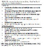
    It is recommended that clinicians pay attention to cer-tain characteristics to improve the accuracy of differen-tial MSA diagnosis 9. Table 3 shows these character-istics recommended for clinically differential diagnosis of MSA.

    Büyütmek İçin Tıklayın
    Table 3: Recent clinical and investigation findings based on clinical judgment to be considered in future.

    The relatively low number of patients included in the study and lack of neuropathological examination of our MSA patients may be accepted as limitations of our study.

  • Başa Dön
  • Özet
  • Giriş
  • Materyal ve Metot
  • Bulgular
  • Tartışma
  • Sonuç
  • Kaynaklar
  • Sonuç
    MSA is frequently diagnosed inaccurately by clinicians examining the patient for the first time, particularly in initial years of the disease. Therefore, clinicians should be careful and try their best to observe red flags while examining patients who may have differential symp-toms associated with MSA.
  • Başa Dön
  • Özet
  • Giriş
  • Materyal ve Metot
  • Bulgular
  • Tartışma
  • Sonuç
  • Kaynaklar
  • Kaynaklar

    1) Stefanova N, Bucke P, Duerr S, Wenning GK. Multiple system atrophy: an update. The Lancet Neurology 2009; 8: 1172-8.

    2) Wenning GK, Ben Shlomo Y, Magalhaes M, Daniel SE, Quinn NP. Clinical features and natural history of multiple system atrophy. An analysis of 100 cases. Brain 1994; 4 : 835-845.

    3) Low PA, Reich SG, Jankovic J, et al. Natural history of multiple system atrophy in the USA: a prospective cohort study. The Lancet Neurology 2015; 7: 710-9.

    4) Gilman S, Wenning GK, Low PA, et al. Second consensus statement on the diagnosis of multiple system atrophy. Neurology 2008; 9: 670-6.

    5) Koga S, Aoki N, Uitti RJ, et al. When DLB, PD, and PSP masquerade as MSA: an autopsy study of 134 patients. Neurology 2015; 5: 404-2.

    6) Osaki Y, Ben-Shlomo Y, Lees AJ, Wenning GK, Quinn NP. A validation exercise on the new consensus criteria for multiple system atrophy. Movement Disorders 2009; 15: 2272-6.

    7) Kim HJ, Stamelou M, Jeon B. Multiple system atrophy-mimicking conditions: Diagnostic challenges. Parkinsonism & Related Disorders. 2016; 22: 12-5.

    8) Wenning GK, Tison F, Seppi K, et al. Development and validation of the Unified Multiple System Atrophy Rating Scale (UMSARS). Movement Disorders 2004; 12: 1391-402.

    9) Kim HJ, Jeon BS, Jellinger KA. Diagnosis and differential diagnosis of MSA: boundary issues. J Neurol 2015; 8: 1801-13.

    10) Ciolli L, Krismer F, Nicoletti F, Wenning GK. An update on the cerebellar subtype of multiple system atrophy. Cerebellum Ataxias 2014; 10: 1-14.

    11) Klockgether T. Sporadic ataxia with adult onset: classification and diagnostic criteria. The Lancet Neurology 2010; 1: 94-104.

    12) Stamelou M, Hoeglinger GU. Atypical parkin-sonism: an update. Curr Opini Neurol 2013; 4: 401-5.

    13) Gatto E, Rodriguez-Violante M, Cosentino C, et al. Pan-American Consortium of Multiple System Atrophy (PANMSA). A Pan-American multicentre cohort study of multiple system atrophy. J Parkinson's Dis 2014; 4: 693-8.

    14) Kollensperger M, Geser F, Ndayisaba JP, et al. Presentation, diagnosis, and management of multiple system atrophy in Europe: final analysis of the European multiple system atrophy registry. Movement Disorders 2010; 15: 2604-12.

    15) Senard JM, Rai S, Lapeyre-Mestre M, et al. Prevalence of orthostatic hypotension in Parkin-son's disease. J Neurol, Neurosurg Psychi 1997; 5: 584-9.

    16) Petrovic IN, Ling H, Asi Y, et al. Multiple system atrophy-parkinsonism with slow progression and prolonged survival: a diagnostic catch. Movement Disorders 2012; 9: 1186-90.

    17) Fanciulli A, Wenning GK. Multiple-system atrophy. New Eng J Med 2015; 14: 1375-6.

    18) Vanacore N, Bonifati V, Fabbrini G, et al. Epidemiology of multiple system atrophy. ESGAP Consortium. European Study Group on Atypical Parkinsonisms. Neurol Sci 2001; 1: 97-9.

    19) Jain RS, Sannegowda RB, Agrawal A, Hemrajani D, Jain R, Mathur T. 'Hot cross bun' sign in a case of cerebrotendinous xanthomatosis: a rare neuroimaging observation. BMJ Case Reports 2013; 14: 1-3.

    20) Lee YC, Liu CS, Wu HM, Wang PS, Chang MH, Soong BW. The 'hot cross bun' sign in the patients with spinocerebellar ataxia. Eur J Neurol 2009; 4: 513-6.

    21) Muqit MM, Mort D, Miskiel KA, Shakir RA. "Hot cross bun" sign in a patient with parkinso-nism secondary to presumed vasculitis. J Neurol, Neurosurg Psychi 2001; 4: 565-6.

    22) Meijer FJ, Aerts MB, Abdo WF, et al. Contribution of routine brain MRI to the differential diagnosis of parkinsonism: a 3-year prospective follow-up study. J Neurol 2012; 5: 929-35.

    23) Ito S, Shirai W, Hattori T. Evaluating poste-rolateral linearization of the putaminal margin with magnetic resonance imaging to diagnose the Parkinson variant of multiple system atrophy. Movement Disorders 2007; 4: 578-81.

  • Başa Dön
  • Özet
  • Giriş
  • Materyal ve Metot
  • Bulgular
  • Tartışma
  • Sonuç
  • Kaynaklar
  • [ Başa Dön ] [ Özet ] [ PDF ] [ Benzer Makaleler ] [ Yazara E-Posta ] [ Editöre E-Posta ]
    [ Ana Sayfa | Editörler | Danışma Kurulu | Dergi Hakkında | İçindekiler | Arşiv | Yayın Arama | Yazarlara Bilgi | E-Posta ]