[ Ana Sayfa | Editörler | Danışma Kurulu | Dergi Hakkında | İçindekiler | Arşiv | Yayın Arama | Yazarlara Bilgi | E-Posta ]
Fırat Tıp Dergisi
2019, Cilt 24, Sayı 1, Sayfa(lar) 018-021
[ Özet ] [ PDF ] [ Benzer Makaleler ] [ Yazara E-Posta ] [ Editöre E-Posta ]
Is Size of the Pre-Operative Annular Tear Measurement a Risk for Early Recurrent Lumbar Disc Herniation?
Erdinç ÖZEK1, Ramazan SARI3, Fatih Han BÖLÜKBAŞI2, Gökalp SİLAV3, Mehmet TÖNGE2, İlhan ELMACI3
1İstanbul Bezmialem Vakıf Üniversitesi, Beyin ve Sinir Cerrahi Anabilim Dalı, İstanbul, Türkiye
2İstanbul Memorial Hastanesi, Beyin ve Sinir Cerrahi Anabilim Dalı, İstanbul, Türkiye
3İstanbul Acıbadem Üniversitesi Hastanesi, Beyin ve Sinir Cerrahi Anabilim Dalı, İstanbul, Türkiye
Keywords: Anulus Fibrozus, Nüks Disk Hernisi, Yırtık, Annulous Fibrousus, Recurrent Disc Herniation, Tear
Summary
Objective: Majority of the patients with a history of unsuccessful lumbar surgery consists of recurrent disc herniation. Although risk factors in recurrent disc herniations were comprehensively examined in the literature, association of annular rupture size with recurrence of lumbar disc hernia was not identified.

Material and Method: Two hundred and seventy patients who were operated in our neurosurgery clinic because of single level lumbar disc herniation between 2012 and 2014 were reviewed retrospectively. During follow-up, 18 patients who were reoperated because of recurrent disc herniation within the first 6 months and 18 patients who were followed at least for 2 years without any recurrence were included to the study. Among 252 patients, 18 patients who are under follow-up at least for 2 years were selected randomly. The radiologist measured the size of the annular rupture by Lumbar MRI retrospectively.

Results: In the statistical analysis, mean annular rupture diameter in the recurrence group was measured as 18.87 (7-28.6 mm; sd 6.29) whereas mean annular rupture diameter in the non-recurrent group was found as 8.82 (5.5-14.5 mm; sd 2.83). Diameter of the annular rupture in the non-recurrence group was found statistically and significantly lower than the recurrence group. (p <0.001, t =3.71)

Conclusion: Although risk of recurrence in lumbar disc hernia depends on many factors, possibility to develop recurrent disc hernia of the patients who have larger annular rupture diameters should be considered as a risk factor.

  • Top
  • Summary
  • Introduction
  • Methods
  • Disscussion
  • References
  • Introduction
    Prevalence of low back pain along life varies between 60 to 90% and annual prevalence is 5%; only 1-3% of low back pain cases are related to lumbar disc herniation. Only a small portion of the patients with lumbar disc herniation needs surgical treatment. The reported prevalence of failed back surgery varies between 8-20% 1,2. Recurrent lumbar disc herniation is one of the most common causes of failed back surgery and occurs in 5-15% of all patients which are treated surgically 3,4. Many different definitions have been described for recurrent disc herniation, such as recurrent disc herniation at the same level and side, contralateral disc herniation at the same level and a new disc herniation in another level. In this study we include the patients with only one level disc herniation and which have recurrent disk herniation at the same level and side.

    Carragee et al 5 reported and classified the intra-operative findings of disc herniation and the relations between annular defects. They divided the patients into four groups; fragment-fissure herniaitions, fragment-defect herniations, fragment-contained and no fragment-contained groups. They have founded that the degree of annular competence after discectomy and the type of herniation appear to have value for the prediction of the recurrence of sciatica, reoperation, and clinical outcome following lumbar discectomy. Risk factors for recurrent disc herniation previously reported in the literature include constitutional weak-ness of the annular tissue, exposure to repetitive lifting or vibration, heavy lifting, advanced age, smoking, the preoperative size and level of the disc herniation, and the appearance of the herniation at the time of surgery 6. The aim of this current study is to evaluate the relation of annular tear size with recurrent disc herniation radiologically.

  • Top
  • Summary
  • Introduction
  • Methods
  • Disscussion
  • References
  • Methods
    Study Design
    Two hundred and seventy patients who were operated due to lumbar disc herniation in our clinic, between November 2012 and December 2014 were reviewed retrospectively with the permission of local ethic committee. Patients were operated via standard microsurgery method by the same surgical team (IE, EO, RS, FB, GS). Eighteen patients were detected with early recurrent disc herniation (Group I). Eighteen patients without recurrent disc herniation (Group II) selected randomly and were included in the study. Group I included the patients who had recurrent disc herniation at the same side and less than 6 months. Group II consisted of the patients with no recurrent disc herniation at the same side or another level within 2 years follow-up.

    Surgical Technique
    All patients were operated in the same center by the same surgical team experienced at least 5 years. In all patients standard microsurgical interlaminar approach has been performed. Partial medial facetectomy has been performed if required and in all patients a rectangular incision of annular fibrosus has been performed to remove protrude or extruded disk fragments using 15 No. blade. Loose nucleus pulposus fragments were removed by disk rongeour although did not force to remove all disk contents.

    Patient Selection
    Two hundred and seventy patients who were operated via single level microdiscectomy procedure were reviewed retrospectively and the patients who had multi-level disc herniations with or without modic degenerations were excluded from study. The 18 patients with recurrent disc herniation occurred in a time period less than 6 months were included to study (Group I). Other 18 patients in control group were selected randomly (Group II). Mean age was 49,77 years in Group I and 46.5 years in Group II. Group I included 10 female and 8 male patients, where the Group II had 8 female and 10 male patients. For the Group I the operated disk herniation level was L4-5 for 12 patients, L5-S1 for 4 patients and L3-L4 for 2 patients. Average recurrence time was 11.6 weeks for Group I. All 36 patients had ordinary lumbar MRI (Sagittal and axial plane for T1 and T2 weighted images) without contrast administration. There were no signs of apparent instability findings before the surgery noted in Group II and I.

    Among 252 patients, 18 patients who are under follow-up at least for 2 years were selected randomly and annular tear diameters were measured by a radiologist (Group II). Size of the annular rupture in Group I, patients who were reoperated because of recurrent disc hernia within the first 6 months, was measured by the same radiologist. Fifteen of the patients in Group I were reoperated via short lumbar segment stabilization with fusion and rediscectomy due to risk of second recurrence. Three of the patients refused the recom-mended surgery.

    Patient Evaluation
    The lumbar MRI examinations of the 36 patients (Group I and Group II) were evaluated by at least ten year experienced radiologist retrospectively. The annular rupture was characterized as a hyperintense area on T2-weighted images and show contrast enhancement on postcontrast MR images radiologically 7.

    In the literature postcontrast T1-weighted images have been found more sensitive than the hiperintensity of the T2-weighted images for detecting annular ruptures 8,9. Unfortunately all preoperative lumbar MRI exami-nations did not have postcontrast images, so we choose the T2-weighted images in order to evaluate the annular rupture.

    All MRI examinations had sagittal and transverse T2-weighted images. The right-left diameter of the annular ruptures, characterised as hyperintense area on T2-weighted images, were measured using both the transvers and sagital images. We measured the diameter on the transverse T2-weighted images by checking the extensions of it on the sagital T2-weighted images (Figure 1, Figure 2).
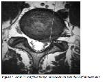

    Click Here to Zoom
    Figure 1: Axial T2 weighted image. Measurement technique of annular tear.
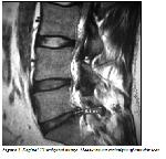

    Click Here to Zoom
    Figure 2: Sagittal T2 weighted image. Measurement technique of annular tear.

    Statistical Analysis
    In the statistical analysis, mean annular rupture diameter in the recurrence group was measured as 18.87 (minimum 7mm-maximum 28.6 mm; sd ±6.29) whereas mean annular rupture diameter in the nonrecurrent group was found as 8.82 (minumum 5.5mmmaximum 14.5mm; sd ±2.83) (Table 1). Using the SPSS statistic soft ware program with Mann Whitney U test the diameter of the annular rupture in the nonrecurrence group was found statistically and significantly lower than the recurrence group (p <0.001).


    Click Here to Zoom
    Table 1: Mean Annular tear diameter of two groups.

  • Top
  • Summary
  • Introduction
  • Methods
  • Disscussion
  • References
  • Discussion
    Recurrent disc herniation rate after lumbar disc surgery was reported as 5-15% in the studies reviewed in the literature although true recurrent disc herniation (at the same level and side) is less than to be thought.

    Annular tear is determined as separation of the annular fibers from their intervertebral insertions. When the tough exterior wall of an intervertebral disc (called the annulus fibrosus) rips, the resulting condition is called an annular tear. Such tears typically occur along the outer edge of a disc, between the layers of the annulus fibrosus, but can also start near the center of the disc and extend all the way to the outside. The extent and positioning of the tear will typically govern the level of pain experienced, which can range from mild to excruciating. In our study diameter of the annular rupture has found to be higher in recurrent group when compared to non-recurrent group (p <0.001). Although there are numerous risk factors have been reported literature for recurrent disc herniation. Carregee et al 5 reported a prospective study that includes intra-operative findings of annulus fibrousus, they divided the findings to four groups such as fragment-fissure herniaitions, fragment-defect herniations, fragment-contained and no fragment-contained groups. They have been reported that the annulus fibrousus competence and the type of the herniaition may indicate recurrent disk herniaition. Ozer et al 10 reported a similar prospective study to Carragee et al in 2013. They have modified the Carrage classification due to low recurrence notes in group I. They have also divided the annular defect in to four types including the intraoperative findings;

    Type I: There is no significant defect on annulus,
    Type II: Annular defect < 4 mm,
    Type III: Annular defect > 4 mm,
    Type IV: Massive-large annular defect.

    Both two studies were based on the intra-operative findings of annular fibrousus so that Ozer et al 11 have been decided intra-operatively to perform posterior percutaneous dynamic transpedicular screwing in Type 2 and 4. Yorimitsu et al 11 have been reported that cases with preserved disk height had favorable early surgical result although a high risk for recurrent disk herniation. This study was contradicted by Cinotti et al 12, and has found that severe disk degeneration increases the risk of recurrent disk herniation, although in this study the case selection was unsuitable because of lesions like narrowed intervertebral foramina, lateral stenosis and epidural scarring.

    Radiological risk factors have been explained detailed by Dora et al 13 recently. Dora et al 13 reported that recurrent disk herniation risk has been increased by degeneration degree of the disc tissue. They have shown that low-grade disk degeneration has an increased risk factor of recurrent disk herniation. They have reached these results by the degree of disk degeneration and the volume of extruded disk herniation, and they did not exclude the patients with Modic Type II and multilevel disk herniations. In the literature it is known that Modic Type II degeneration is related with instability and has higher risk of recurrence. In this study we excluded the patients who had multilevel disk herniations and who had Modic degenerations in both groups. A potential limitation of this study was the small number of cases and the preoperative non-contrast Lumbar MR images. And in control group; the patients were not followed by lumbar MRI, however all patients in control group were checked by phone if they have any complaints or not. One additional limitation was that the patients were operated by the same surgical team, but by not the same surgeon, however using the same surgical technique. Future experimental and/or prospective studies will give us more accurate and detailed information about recurrence of lumbar disk herniations. Although risk of recurrence in lumbar disc herniation depends on many factors, possibility to develop recurrent disc hernia of the patients who have larger annular rupture diameters should be considered as a risk factor.

    All authors thanks to Dr. Özdil Başkan for her contribution.

  • Top
  • Summary
  • Introduction
  • Methods
  • Discussion
  • References
  • References

    1) Crock HV. Observations on the management of failed spinal operations. J Bone Joint Surg Br 1976; 58: 193-9.

    2) O’Sullivan MG, Connolly AE, Buckley TF. Recurrent lumbar disc protrusion. Br J Neurosurg 1990; 4: 319 -25.

    3) Gaston P, Marshall RW. Survival analysis is a better estimate of recurrent disc herniation. J Bone Joint Surg Br 2003; 85: 535‐7.

    4) Suk KS, Lee HM, Moon SH, Kim NH. Recurrent lumbar disc herniation: Results of operative management. Spine (Phila Pa 1976) 2001; 26: 672‐6.

    5) Carragee EJ, Han MY, Suen PW, Kim D. Clinical outcomes after lumbar discectomy for sciatica: the effects of fragment type and anular competence. J Bone Joint Surg Am 2003; 85: 102-8.

    6) Shimia M, Babaei-Ghazani A, Sadat BE, Habibi B, Habibzadeh A. Risk factors of recurrent lumbar disk herniation. Asian J Neurosurg 2013; 8: 93-6.

    7) Weishaupt D, Zanetti M, Hodler J, Boos N. MR imaging of the lumbar spine: prevalence of intervertebral disk extrusion and sequestration, nerve root compression, end plate abnormalities, and osteoarthritis of the facet joints in asymptomatic volunteers. Radiology 1998; 209: 661-6.

    8) Ross JS, Modic MT, Masaryk TJ. Tears of the anulus fibrosus: assessment with Gd-DTPA-enhanced MR imaging. AJNR Am J Neuroradiol 1989; 10: 1251–4.

    9) Stadnik TW, Lee RR, Coen HL, Neirynck EC, Buisseret TS, Osteaux MC. Annular tears and disk herniation: prevalence and contrast enhancement on MR images in the absence of low back pain or sciatica. Radiology 1998; 206: 49–55.

    10) Ozer AF, Keskin F, Oktenoglu T et al. Anovel approach to the surgical treatment of lumbar disc herniations: indications of simple discectomy and posterior transpedicular dynamic stabilization based on carragee classification. Adv Orthop 2013; 2013: 270-565.

    11) Yorimitsu E, Chiba K, Toyama Y, Hirabayashi K. Long-term outcomes of standard discectomy for lumbar disc herniation: a follow-up study of more than 10 years. Spine (Phila Pa 1976) 2001; 15; 26: 652-7.

    12) Cinotti G, Gumina S, Giannicola G, Postacchini F. Contralateral recurrent lumbar disc herniation. Results of discectomy compared with those in primary herniation. Spine (Phila Pa 1976) 1999: 15; 24: 800-6.

    13) Dora C, Schmid MR, Elfering A, Zanetti M, Hodler J, Boos N. Lumbar disk herniation: do MR imaging findings predict recurrence after surgical diskectomy? Radiology 2005; 235: 562-7.

  • Top
  • Summary
  • Introduction
  • Methods
  • Discussion
  • References
  • [ Başa Dön ] [ Özet ] [ PDF ] [ Benzer Makaleler ] [ Yazara E-Posta ] [ Editöre E-Posta ]
    [ Ana Sayfa | Editörler | Danışma Kurulu | Dergi Hakkında | İçindekiler | Arşiv | Yayın Arama | Yazarlara Bilgi | E-Posta ]