 |
 |
| [ Ana Sayfa | Editörler | Danışma Kurulu | Dergi Hakkında | İçindekiler | Arşiv | Yayın Arama | Yazarlara Bilgi | E-Posta ] | |
| Fırat Tıp Dergisi | |||||
| 2007, Cilt 12, Sayı 2, Sayfa(lar) 151-153 | |||||
| [ Özet ] [ PDF ] [ Benzer Makaleler ] [ Yazara E-Posta ] [ Editöre E-Posta ] | |||||
| Left Inferior Vena Cava Associated with Nutcracker Phenomenon | |||||
| Şerife ULUSAN, Zafer KOÇ | |||||
| Başkent Üniversitesi, Adana Uygulama ve Araştırma Merkezi Radyoloji Anabilim Dalı, ADANA | |||||
| Keywords: Left inferior vena cava, nutcracker phenomenon, Sol inferior Vena Kava, Nutcracker Fenomeni | |||||
| Summary | |||||
Left inferior vena cava (LIVC) is a congenital vascular malformation characterized by crossing over to the right via the left renal vein or more
inferiorly crossover. The nutcracker phenomenon is defined as entrapment of the left renal vein between the aorta and superior mesenteric artery. The
association between the nutcracker phenomenon and LIVC has not been reported previously. We report the radiologic and clinical findings of LIVC
and nutcracker phenomenon. ©2007, Fırat Üniversitesi, Tıp Fakültesi |
|||||
| Introduction | |||||
The nutcracker phenomenon is rare but well acknowledged
cause of hematuria, ureteral and peripelvic varices, and
unexplained left flank and abdominal pain 1-3. Varying
degrees of proteinuria have been observed in some patients
with the nutcracker phenomenon 4,5. Left IVC has been well-described and occurs in 0.2%-0.5 % of the general population 6. In most cases via the left renal vein or more inferiorly, and crossover is usually anterior to the aorta 6,7. In this study, we present a case of LIVC associated with nutcracker phenomenon. |
|||||
| Case Presentation | |||||
An 80-year-old woman was admitted to our hospital in
February 2004 with complaints of abdominal pain. She had a
palpable mass in the right upper quadrant and was referred to
our unit for radiologic evaluation. Urinalysis showed 1+
hematuria and proteinuria 0.30 g/L (normal range, 0.03-0.14
g/L). Serum creatinine, blood urea nitrogen, total protein,
albumin, and creatinine clearance levels were within normal
limits. Her white blood cell count was 45.300 K/mm3 (normal
range 4500-10000), and her platelet count was 85.300 K/mm3
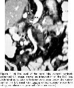
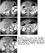
(normal range 130.000-400.000). The patient was evaluated by abdominal ultrasound and CT examinations. Abdominal ultrasound revealed a 16 cm x 11 cm x 11 cm heterogeneous/hyperechoic mass in the left lobe of the liver. The biliary tree was not dilated. Noncontrast and contrast-enhanced abdominal and pelvic CT scans showed the mass had invaded the celiac truncus and the head of the pancreas and, the liver was not cirrhotic. A Tru-cut biopsy was performed, and the microscopic findings suggested a metastatic neoplasm originating from an adenocarcinoma. Multiplanar contrast-enhanced CT of the abdomen also showed the interposition of the IVC (Figure 1). At the level of the renal hila; the left vena cava passes either in front of the aorta or runs to right as a left renal vein and becomes stenosed and the distal portion of the left renal vein was dilated. The criterion for diagnosis of nutcracker phenomenon is accepted the ratio of anteroposterior diameter of the stenosed segment (between the aorta and SMA) to the left renal vein near the hilum is more than 1/5 (8). In our case, the ratio of stenosed segment of the left renal vein (between the aorta and SMA) and dilated segment of the left renal vein near the hilum of the anteroposterior diameters was measured the approximetly 2mm/12mm. Consecutive axial CT images showed compression of the left renal vein between the aorta and the superior mesenteric artery (SMA) (Figure 2).
Owing to her poor clinical state, she did not operate. Twenty days later, the patient died. |
|||||
| Discussion | |||||
The left renal vein is normally located between the abdominal
aorta and the superior mesenteric artery (SMA). In the
nutcracker phenomenon, there is an abnormal branching of the
SMA from the abdominal aorta 1. Subsequently, the left renal
vein is compressed between these arteries. This compression
causes elevated pressure in the left renal vein and this high
pressure then result in rupture of the thin-walled veins of the
renal collecting system. Permanent left renal vein hypertension
may affect collateral veins causing dilatation of the gonadal
vein and varicocele 1-3. Enhanced CT of the abdomen,
sagittal magnetic resonance angiography, and left renal
venography are helpful in establishing diagnosis of the
nutcracker phenomenon 4. Usual clinical presentation of the nutcracker phenomenon may include flank pain or abdominal back pain only 1-3. Our patient presented with abdominal pain and hematuria. Due to poor health condition with metastatic liver disease, she was not evaluated for nutcracker phenomenon. In the fifth week of gestation, 3 pairs of major veins differentiate: the vitelline veins, the umbilical veins, and the cardinal veins. From a division of the cardinal veins, the subcardinal veins and the lower portion of the IVC form 6. The subcardinal system is composed of paired veins on either side of the developing abdominal aorta. The left subcardinal vein typically regresses to form the left gonadal vein and the left renal vein 6,7. If this left subcardinal venous system does not regress and the left renal vein remains, an LIVC, also called a transposed IVC is formed. The most common variations are duplicate IVC, retroaortic renal veins, and circumaortic venous rings 6,7. However, LIVCs are rare, with a reported incidence of only 0.2%-0.5% 6,7. The left IVC is of no importance unless a surgical operation is being considered. Its surgical implications can be important because it may complicate the surgery of aneurysms of the abdominal aorta. To the best of our knowledge, this is the first reported case of an association between of LIVC and the nutcracker phenomenon. We believe that LIVC and the nutcracker phenomenon may occur more frequently than has been reported owing to their anatomic relationship. |
|||||
| References | |||||
1) Cuellar I Calabria H, Quiroga et al. A. Nutcracker or left renal
vein compression phenomenon: multidetector computed
tomography findings and clinical significance. Eur Radiol. 2005
15:1745-1751.
2) Chang CT, Hung CC, Ng KK, et al Nutcracker syndrome and left
unilateral haematuria. Nephrol Dial Transplant. 2005; 20: 460-
3) Kaneko K, Kiya K, Nishimura K, et al. Nutcracker phenomenon
demonstrated by three-dimensional computed tomography.
Pediatr Nephrol. 2001; 16: 745-747.
4) Ekim M, Bakkaloglu SA, Tumer N, et al Orthostatic proteinuria
as a result of venous compression (nutcracker phenomenon) a
hypothesis testable with modern imaging techniques. Nephrol
Dial Transplant. 1999; 14:826-827.
5) Kaneko K, Ohtomo Y, Yamashiro Y, et al Magnetic resonance
angiography in nutcracker phenomenon. Clin Nephrol. 1999; 51:
259-260.
6) Minniti S, Visentini S, Procacci C. Congenital anomalies of the
venae cavae: embryological origin, imaging features and report of
three new variants. Eur Radiol. 2002; 12: 2040-2055.
|
|||||
| [ Başa Dön ] [ Özet ] [ PDF ] [ Benzer Makaleler ] [ Yazara E-Posta ] [ Editöre E-Posta ] | |||||
| [ Ana Sayfa | Editörler | Danışma Kurulu | Dergi Hakkında | İçindekiler | Arşiv | Yayın Arama | Yazarlara Bilgi | E-Posta ] |

