 |
 |
| [ Ana Sayfa | Editörler | Danışma Kurulu | Dergi Hakkında | İçindekiler | Arşiv | Yayın Arama | Yazarlara Bilgi | E-Posta ] | |
| Fırat Tıp Dergisi | |||||||||||||
| 2008, Cilt 13, Sayı 2, Sayfa(lar) 150-152 | |||||||||||||
| [ Özet ] [ PDF ] [ Benzer Makaleler ] [ Yazara E-Posta ] [ Editöre E-Posta ] | |||||||||||||
| Immature Mediastinal Teratoma: Radiological Findings (A Case Report) | |||||||||||||
| Ayse Murat AYDIN, Mustafa KOÇ, A.Kursad POYRAZ | |||||||||||||
| Fırat Üniversitesi Tıp Fakültesi Radyoloji Anabilim Dalı, ELAZIĞ | |||||||||||||
| Keywords: Mediastinum, Immature teratoma, Computerized tomography, Magnetic resonance imaging, Mediasten, İmmatür teratom, Bilgisayarlı tomografi, Manyetik rezonans görüntüleme | |||||||||||||
| Summary | |||||||||||||
Immature mediastinal teratomas are rare malignant neoplasms that can metastasize and recur, are characterized by the presence of various tissues that histologically resemble embryonal structures. Computerized tomography (CT) shows the location and extent of the tumors as well as intrinsic elements including soft tissue, fat, fluid, and calcification. CT is the radiological modality choice for the diagnostic evaluation of these tumors and also combination of magnetic resonance imaging (MRI) best defines the characterisation, location and operability of tumor. In this report, we present radiological findings of immature mediastinal teratoma.©2008, Fırat Üniversitesi, Tıp Fakültesi |
|||||||||||||
| Introduction | |||||||||||||
Mature and immature teratomas are the basic pathologic types of teratomas, which mostly occur in the mediastinum and pericardium. Mediastinal teratomas have been classified as mature when there is histologically well differantiated tissue and as immature when they contain so-called immature epithelial and mesenchymal elements as well as mature tissue (especially tissues of neuroepithelia)1,2. Immature mediastinal teratomas are rare, found in only 1% of all mediastinal teratomas3. Immature mediastinal teratomas can metastasize and recur and are characterized by the presence of various tissues that histologically resemble embryonal structures3,4. In the literature there is no detail about radiological images of this lesion enough. The combination of CT and MRI best defines the location and operability of tumor. In this report, we describe radiological findings of a rare case of immature mediastinal teratoma arising from the right anterior mediastinum. |
|||||||||||||
| Case Presentation | |||||||||||||
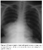
A 26-year-old male patient was admitted to our hospital with complaints of cough and dyspnea for 4 months. He was also complainig from sputum production, hemoptysis and right chest pain for 20 days. Night fever and perspiration was accompanying symptoms. Physical examination revealed decreased breath sounds in lower zone of right lung. Routine laboratory tests was within normal limit. Posteroanterior chest radiograph revealed a large, smooth edged opasity between right pulmonary hilus and right diaphragma. Opacity was blurring the contours of diaphragma and heart (Figure 1).
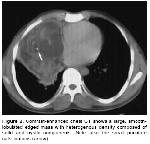
Contrast-enhanced chest CT showed a smooth-lobulated edged mass with heterogenous density composed of solid and cystic components (density values mostly ranged from 80 to 110 HU at the more hypodense area and 10 HU at the cystic region), which located between anterior mediastinum and right pleura and right diaphragma. Mass were also containing small punctate calsifications (density value was approximately 300 HU) and extending to anterior chest wall (Figure 2).
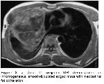
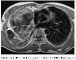
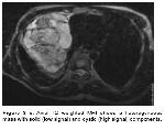
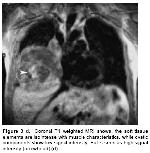
The mass was obliterating perivascular and pericardial fat planes. Right lungs volume was decreased. Anterior segment of upper lobe, middle lobe and mediobasal segment of lower lobe of right lung parenchyme was obliterated. Chest MRI showed a mass which located within anterior mediastinum and right hemithorax with dimensions of 10x12x10 cm. Mass was smooth-lobulated edged with heterogenous intensity on both T1 and T2 weighted images and also some areas with high signal intensity on T1 weighted images which demonstrate fat within the mass. Post-contrast T1 weighted images showed, mass have enhanced capsule and solid components. Mediastinal and chest wall fat planes were obliterated. There was an irregularity at the posterior contour of the mass and strong enhancement at the adjacent lung parenchyme due to inflamation (Figure 3a-d).
At operation, a median sternotomy was used to approach mass, which was totally removed. Histopathology proved it to be an immature teratoma. |
|||||||||||||
| Discussion | |||||||||||||
Immature teratomas are rare tumors that differ from benign teratomas in that the component tissue resembles that observed in the fetus or embryo. Any type of tissue may be represented in immature teratoma, the main component is usually neurogenic, but mesodermal elements are also common. Immature teratomas grow rapidly and frequently penetrate the capsule with spread or metastases4,5. The pathogenesis of extragonadal immature teratoma is not completely understood. Clinically it is not possible to differentiate mature teratoma from immature teratoma. The differentiation can only be made by careful histological examination4. Crosssectional imaging using CT, MR imaging, allows identification of different elements within these tumors including soft tissue, fluid, fat and calcium. Mediastinal immature teratoma typically manifests on CT as a heterogeneous anterior mediastinal mass containing soft-tissue, fluid, fat, or calcium attenuation, or any combination of the four. Fluid-containing cystic areas, fat, and calcification occur frequently. Cystic lesions without fat or calcium were seen in 15 % of tumors. Fat-fluid levels, considered highly specific for the diagnosis of mediastinal mature teratoma, are uncommon. CT is the imaging technique of choice in the evaluation of these lesions. CT is also useful in the evaluation of associated pulmonary opacities, which usually represent atelectasis, pneumonitis, or both6. The most common MRI appearance of a teratoma is that a heterogeneous anterior mediastinal mass. The soft tissue elements are isointense with muscle characteristics, while cystic components show low signal intensity on T1-weighted images and high signal on T2-weighted images. Visualization of fat is, high signal intensity on T1-weighted images that useful in determining the diagnosis. MRI is also useful in the evaluation of the inflammation around the tumor7. The differential diagnosis of fat-containing mediastinal masses includes mediastinal fat pad, mediastinal lipomatosis, lipoma, omental herniation, thymolipoma, and liposarcoma. However, immature teratomas are easily distinguished from these lesions by their cystic appearance with predominance of fluid elements and high frequency of calcification, findings readily seen on CT scans. In our case also, like the literature, a heterogeneous smooth-lobulated edged mass was seen arising from the right anterior mediastinum containing soft-tissue, fluid, fat and calcium attenuation. In patients >15 years of age with immature mediastinal teratoma, complete surgical resection of the tumour is indispensable. When complete resection is combined with chemotherapy, long survival can be expected. Conversely, unless both treatment methods are performed, the outcome is very poor3. Ozeki et al.8 concluded that radiotherapy should be added to combination of intensive chemotherapy and surgical resection in immature mediastinal teratomas treatment. In conclusion, immature teratomas are rare tumors that differ from benign teratomas. The differentiation can only be made by careful histological examination. CT is the radiological modality choice for the diagnostic evaluation of these tumors and also combination of MRI best defines the characterisation, location and operability of tumor. |
|||||||||||||
| References | |||||||||||||
1) Aksoy F, Sen C, Danisment N. Congenital mediastinal immature teratoma: a case report with autopsy findings. Turk J Pediatr 2002; 44:76-79.
2) Moran CA, Suster S. Primary germ cell tumors of the mediastinum: II. Mediastinal seminomas-a clinicopathologic and immunohistochemical study of 120 cases. Cancer 1997; 80:691-698.
3) Das BB, Richardson C, Lala R, Sahoo S. Primary Immature Mediastinal Teratoma in a Newborn Indian Pediatrics 2001; 38:1181-1184.
4) Col C. Immature teratoma in both mediastinum and liver of a 21-Year-old female patient. Acta Med Austriaca 2003; 30:26-28.
5) Hiroshima K, Toyozaki T, Iyoda A, et al. Apoptosis and proliferative activity in mature and immature teratomas of the mediastinum. Cancer 2001; 92:1798-1806.
6) Choi SJ, Lee JS, Song KS, Lim TH. Mediastinal teratoma: CT differentiation of ruptured and unruptured tumors. AJR Am J Roentgenol 1998; 171:591-594.
|
|||||||||||||
| [ Başa Dön ] [ Özet ] [ PDF ] [ Benzer Makaleler ] [ Yazara E-Posta ] [ Editöre E-Posta ] | |||||||||||||
| [ Ana Sayfa | Editörler | Danışma Kurulu | Dergi Hakkında | İçindekiler | Arşiv | Yayın Arama | Yazarlara Bilgi | E-Posta ] |
